Schematic

CXI Schematic

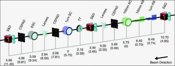
CXI Beamline Sections

Acronym Full Name Devices Purpose
XRT:DIA DIAgnostics stand in the X-ray Tunnel
  • Pulse Picker
  • Attenuators (10 filters)
  • X-ray Focusing Lenses
  • Rapidly shutter the X-ray beam
  • Control the incident beam intensity
  • Pre-focus the X-ray beam or produce a ~10 micron focus at CXI
DG1 DiaGnostics stand 1
  • Reference Laser
  • Slits 1
  • Profile-Intensity Monitor
  • Differential Pump
  • Produce a visible reference line for alignment
  • Slit the beam down and reduce unwanted halo, Define the beam entering the KB mirrors
  • Measure the incidnet beam profile entering the CXI hutch
  • Separate the vacuum of the KB mirrors from the upstream beamline
KB1 1 micron KB mirrors
  • Slits 2
  • 1 micron KB mirrors
  • Slits 3
  • Slit the beam down and clean the beam before entering the KB mirrors
  • Focus the X-ray beam to a ~1 micron spot
  • Slit the beam down and clean the beam entering the KB01 mirrors or the beam exiting the KB1 mirrors
KB01 or KB2 0.1 micron KB mirrors
  • 100 nm KB mirrors
  • Focus the X-ray beam to a ~100 nm spot
SC01 or SC2 0.1 micron Sample Chamber
  • 100 nm interaction region
  • Location of the ~100 nm focus
DSA Detector Stage (A location)
  • Full size CSPAD
  • X-ray Focusing Lenses
  • Attenuator or beam stop
  • Forward scattering measurements with the nanofocus chamber
  • Refocusing or collimating the 100 nm focus for reuse downstream
  • Attenuate the beam passing through the CSPAD hole or block it entirely
DG2 DiaGnostics stand 2
  • CSPAD 140K or X-ray Focusing Lenses
  • Slits 4
  • Intensity-Position Monitor
  • Profile-Intensity Monitor
  • Differential Pump
  • Measure the low angle forward scattering from the 100 interaction region

OR

  • Change the divergence of the 1 micron KB beam and control the spot size in SC1
  • Slit the beam down after the refociusing lenses or clean the beam before after the 1 micron KB mirrors
  • Non-destructive relative measurement of the pulse intensity
  • Measure the beam profile on the way to the 1 micron focus, after the 100 nm focus or after the refocusing lenses
  • Isolate the 1 micron sample chamber environment from the upstream part of the beamline
DSB Detector Stage (B location)
  • Full size CSPAD

OR

  • Time Tool
  • Pulse Selector
  • Attenuators (6 filters)
  • Slits 5
  • Differential Pump
  • Back scattering measurements with the 1 micron focus

OR

  • Cross-correlate the arrival time of the X-rays and pump lasers
  • Rapidly shutter the X-ray beam or control the repetition rate for SC1
  • Control the incident beam intensity
  • Slit the beam down after the refocusing lenses or clean the beam before after the 1 micron KB mirrors
  • Isolate the 1 micron sample chamber environment from the upstream part of the beamline
SC1 1 micro Sample Chamber
  • 1 micron interaction region
  • Location of the ~1 micron focus and of the refocused 100 nm beam
DSC Detector Stage (C location)
  • Full size CSPAD (Front detector for SC1)
  • Attenuator or beam stop

Coming Soon

  • Pulse Selector for refocused 1 micron beam
  • X-ray focusing lenses for refocusing or collimating the 1 micron focus
  • Forward scattering measurements with the micron focus chamber (High angle detector)
  • Attenuate the beam passing through the CSPAD hole or block it entirely

Coming Soon

  • Rapidly shutter the X-ray beam or control the repetition rate for SSC
  • Refocusing or collimating the 1 micron focus for reuse downstream
SSC or SC3 Serial Sample Chamber
  • Interaction for refocused 1 micron beam
  • Location of the refocused 1 micron beam
DSD Detector Stage (D location)
  • Full size CSPAD (Back detector for SC1)

OR

  • Full size CSPAD (Detector for SSC)
  • Attenuator or beam stop
  • Forward scattering measurements with the micron focus chamber (Low angle detector)

OR

  • Forward scattering measurements with the refocused beam in SSC
  • Attenuate the beam passing through the CSPAD hole or block it entirely
DG3 DiaGnostics stand 3
  • Single Shot Spectrometer
  • Measure the single shot X-ray spectrum
DG4 DiaGnostics stand 4
  • Intensity-Position Monitor
  • Profile-Intensity Monitor
  • Non-destructive relative measurement of the pulse intensity
  • Measure the beam profile at the end of the CXI hutch, after all focusing optics and all interactions with samples or diagnostics